The aging spine
By Carlo Ammendolia
Features Clinical TechniquesManaging degenerative lumbar spinal stenosis in older adults
 Lumbar spinal stenosis is a leading cause of pain and disability among older adults. Pain in the lower extremities is one of the symptoms.
 A 70-year-old male presents to your clinic complaining of worsening back pain and lower extremity pain, numbness, heaviness and weakness. He was a regular walker, walking up to four kilometers per day, and now he cannot walk more than a half block and has to sit down for relief. He has high blood pressure but is otherwise healthy.
How would you assess and manage this patient?
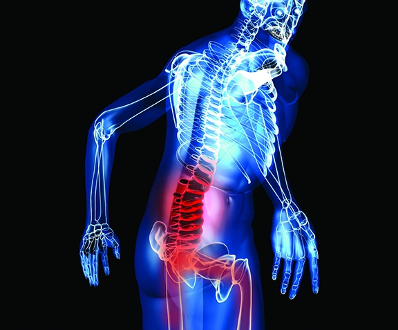
Degenerative lumbar spinal stenosis (DLSS) is a leading cause of pain, disability and loss of independence in older adults. Lumbar spinal stenosis refers to a focal narrowing of the central canal and/or lateral foramina of the spine, usually identified by imaging. Evidence of narrowing of the spinal canals alone without clinical information is not meaningful since 30 per cent of patients over the age of 55 have moderate spinal stenosis and have no symptoms.
Neurogenic claudication is the term used to describe the clinical syndrome caused by lumbar spinal stenosis. It is characterized by bilateral or unilateral buttock, lower extremity pain, heaviness, numbness, tingling or weakness, precipitated by walking and standing, and relieved by sitting and bending forward.
There are many causes of narrowing of the spinal canals. These include congenitally narrowed pedicles, spondylolisthesis, metabolic bone diseases – like Paget’s disease and acromegaly – and previous spine surgery which can cause reactive bone formation within the canals. By far the most common cause of spinal stenosis is degenerative arthritis.
Degenerative arthritis or osteoarthritis is a wear-and-tear type of arthritis that we all get to some degree when we age. Degenerative arthritis results in thinning and bulging of the intervertebral discs, thickening of the facet joints, and infolding and thickening of the internal spinal ligaments. Degenerative spinal changes lead to a decrease in the cross sectional area of the spinal canals and potential compression to the spinal nerves that travel to the lower extremities. The narrowed spinal canals also restrict blood flow to the spinal nerves, which needs oxygen to function. This leads to neuro-ischemia and hypoxia to the nerves which result in lower extremity pain and claudication.
Limited walking ability is the dominant functional impairment caused by symptomatic DLSS. Inability to walk among individuals with symptomatic DLSS leads to a sedentary lifestyle and a progressive decline in health status. DLSS is a chronic disease that can deteriorate with age.
Reliable data on the prevalence of symptomatic DLSS is lacking. A Japanese study revealed that almost 50 per cent of patients who present themselves to a primary care doctor with numbness and tingling of the lower extremities have neurogenic claudication due to lumbar spinal stenosis, with an average age of 65 years for females and 55 for males.
An interesting phenomena associated with DLSS is the dynamic nature of the symptoms. Symptoms are usually precipitated by standing and walking. The longer one stands or walks the more intense the symptoms become. However, sitting or stooping forward or lying down results in a rapid reduction or elimination of the symptoms. This is explained by the change in the cross sectional area of spinal canals with changing posture. Lumbar flexion, which occurs when you sit or stoop forward, increases the spinal cross sectional area, reduces spinal nerve compression and restores spinal blood supply. Whereas, lumbar extension (we tend to maintain a lumbar lordosis when we stand and walk) decreases the cross sectional area and increases nerve pressure and symptoms.
The patient described at the beginning of this article demonstrates this typical phenomenon and, at 70 years old, likely has degenerative changes in the spine. Patients with DLSS will typically describe their leg symptoms as numbness, tingling, pins and needles, weakness or heaviness in the buttock, posterior thigh and lower leg that can impact their ability to walk. Back pain can be present, but not always, and can follow the same dynamic pattern.
Physical examination
On physical examination, patients with DLSS tend to stand with a stooped posture. Range of motion testing typically demonstrates little difficulty during forward flexion. However, lumbar extension is usually limited and painful, and can sometimes reproduce lower extremity symptoms. Lumbar extension position may have to be maintained for a period of time before leg symptoms are reproduced.
Balance testing may reveal difficulty and many patients will use a cane for added security. Heel-toe walking and a standing squat may demonstrate weakness that reflects involvement of a specific nerve root(s). However, this is usually seen in more long standing DLSS. The same holds true for sensory deficits, which will follow a dermatomal pattern. Deep tendon reflexes of the lower extremities are often difficult to elicit in older patients, although this is usually noted symmetrically.
Supine straight leg raising is usually negative in DLSS because this maneuver introduces lumbar flexion and reduces nerve compression. Loss of muscle tone and, in more advanced cases, muscle atrophy are noted due to nerve compression and disuse (as a result of a more sedentary lifestyle). It is important to assess the lower extremity pulses to ensure they are present and symmetrically equal.
Differential diagnosis
One of the key goals in assessment is to determine the source of the patient’s symptoms and functional limitations in order to provide appropriate treatment.
Osteoarthritis of the hip is highly prevalent in this patient population and can mimic DLSS. Symptoms are usually located in the back, buttock and groin, and pain can be referred distally to the knee. The symptoms are worse standing and walking and relieved by sitting and lying down. However, there tends to be a characteristic limping gait with a lateral shift in the pelvis on the involved side when weight bearing (Trendelenberg Sign) and usually no large change in symptoms when walking with a stooped posture or when using a shopping cart. A hip exam is critical and often demonstrates painful and restricted internal rotation and flexion during range of motion testing. This testing usually reproduces the patient’s symptoms. Hip-spine syndrome is the diagnosis given when imaging confirmed osteoarthritis of the hip and DLSS are present at the same time, and both contribute to the patient’s symptoms and functional limitations.
Vascular claudication is a condition that impacts walking ability and also highly present in older patients. It is usually a result of peripheral arterial disease (PVD) – the most common being due to atherosclerosis, which impacts blood flow to the lower extremities, especially the leg muscles. Like DLSS, symptoms progress with walking and relieved by rest, but unlike DLSS stooped posture while walking does not change symptoms or ability to walk. There can be trophic discolouration of skin noted in the lower extremities, especially involving the distal leg and foot. Diminished or absent pulses of the lower extremities can be noted but often not reliable for diagnosis.
An arterial Doppler test is recommended to more accurately assess blood flow. About 30 per cent of DLSS patients also have confirmed PVD, which again makes diagnosis a challenge. Other conditions in the differential diagnosis of symptomatic DLSS include trochanteric bursitis, diabetic neuropathy and meralgia paresthetica. Disc herniation is more frequent in the 30 to 40 age groups and are usually associated with painful and restricted forward flexion, a positive straight leg raise and worsening symptoms with sitting, all of which are the antithesis of what is seen with DLSS.
Neurogenic claudication is a clinical diagnosis based on the history and physical examination. Imaging is not necessary. Imaging is needed when red flags are present that suggest other potential serious diseases or conditions such as cancer, infection or spine fracture. Imaging is also needed when the patient’s condition is not improving and there may be the need for surgery or other invasive treatments.
Treatment
DLSS is the most common reason for spine surgery in individuals over the age of 65. There are generally two types of surgery: direct and indirect decompression. Direct decompression involves removal of bony elements within the spine to increase the cross sectional area of the spinal canals. Indirect decompression involves the placement of a device within the spine to change the alignment in an attempt to increase the cross sectional area of the spinal canals. Patients who have leg dominant rather than back dominant symptoms tend to do better after surgery; however, the benefits tend to diminish over time.
The vast majority of individuals with DLSS receive non-surgical care. However, what constitutes effective non-surgical care is unknown. Most common non-surgical therapies include physical therapy, chiropractic, acupuncture, massage therapy, medication and epidural injections. Anti-inflammatory medication and epidural cortisone injections tend to be ineffective since the symptoms are generally due to neuro-ischemia and not inflammation. This is demonstrated by the immediate reduction in symptoms when flexing forward (which restores spinal blood flow). This would not likely be the case if the condition was due to an inflammatory process.
The ability to reduce symptoms of DLSS, by changing spinal posture/structural alignment and/or increasing blood flow to the spinal nerve, provides potential mechanisms for interventions to improve symptoms and walking ability.
The Rebecca MacDonald Centre for Arthritis and Auto-immune Diseases at Toronto’s Mount Sinai Hospital has designed and implemented a six-week self-management training program – The Boot Camp Program – for DLSS. The goal of the training program is to provide patients with the knowledge, skills, self-confidence and physical capacity to manage their symptoms and maximize their function on their own. The program is multimodal, tailored and directed to the structural, functional, physiological and psychosocial consequences of DLSS. It includes structured aerobic exercise (walking, cycling or swimming) to build stamina, self-confidence, lower extremity strength, balance and overall fitness. There is instruction on specific exercises that help build core strength aimed at enabling individuals to reduce the lordosis (body realignment using pelvic tilt) during standing and walking. Education is provided using a cognitive behavioural approach, and is aimed at problem solving, goal setting and building self-confidence to self-manage and maximize mobility. Manual therapy is provided with the aim of improving intersegmental lumbar spine flexion and overall flexibility, with the goal of facilitating the ability to self-align the lumbar spine to a more functional position using the pelvic tilt while standing and walking.
The program is delivered one-on-one over six weeks. During this period, a step-by-step home exercise program is designed and tailored to the individual with the goal of being maintained for life. There are currently two clinical trials evaluating the effectiveness of this approach – one at the University of Toronto and the other at the University of Pittsburgh.
Dr. Carlo Ammendolia, DC, PhD, is the director of the Chiropractic Spine Clinic and the Spinal Stenosis Program at the Rebecca MacDonald Centre for Arthritis and Auto-immune Diseases at Mount Sinai Hospital. He is an assistant professor in the department of health policy, management and evaluation at the University of Toronto. In 2012, Ammendolia was awarded the Professorship in Spine Award from the department of surgery in the faculty of medicine.
Print this page