Editor’s Note: In commemoration of Canadian Chiropractor’s 20th anniversary, we’re bringing back this well-loved column that forms part of this magazine’s history. We asked Dr. Marshall Deltoff to be part of our anniversary issue for old time’s sake.
Admittedly, I used to be an opponent of digital x-rays. I would cringe when a little bubble-wrap filled envelope arrived on my desk, and would quickly bury it under all of the other work I had to do that day. Why? Well, it would take me literally three times longer to read an x-ray CD rather than take films out of an envelope and put them up on the view box.
Each series from the various facilities seemed to have its own unique and complicated “user-friendly” program. I couldn’t, and didn’t want to, take the time to figure them out. When x-ray studies would come into my office for me to report on, I would rush to open the film mailing envelopes first, always leaving the digital ones until the end, and approach those discs with the disdain I felt they deserved as tedious time-wasters.
Since then I must say that I have had a 180-degree change of attitude and in fact, have developed a virtual paradigm shift toward a whole-hearted endorsement of digital radiography in its current form. This happened because, ultimately, I am a scientist and try to be intellectually honest with myself.
Digital radiography has truly matured from its infancy as a “cool, hi-tech, expensive toy” to “must-have technology.” Was the shift difficult for me? For sure. Engrained, longstanding attitudes and beliefs are always kind of hard to change. I was pretty “old school.”
I was taught at CMCC how to hand-tank films as well as use a processor. Culturally, as a radiologist, I liked seeing the film come out of the processor and holding it up to the view box, pleased that I got a good shot. I liked taking my special x-ray marking pencil and ruler and “analyzing” the film with my lines and angles. Holding the film up to the light or the view box was part of what I did, part of my routine, part of my image as an imaging specialist. I handed the x-ray jacket full of x-rays and my report to the eager and anxious patient as part of the expected, sacred ritual of the radiologist. Well, so much for that. Put that diorama next to the stegosaurus skeleton in the museum.
With digital technology, I can, and do, read x-rays for chiropractors from all over the world. A DC can allow me to log on to his/her x-ray lab website, and read the films directly. I can then send a PDF report that doctors can print for their patient file. They can extract the DCM image files from a disc, and put them in my Dropbox – again, allowing quick access to films from anywhere at anytime, and they get their report the same day or next day. Finally, even regular films can be photographed and e-mailed to me as JPEG files for consultation with regard to trauma, arthritis or pathology.
Before my digital conversion I had what I thought were legitimate concerns about patient dosage – but I was labouring under misinformation. I had read that there was significantly more patient dose with digital imaging. But this is 2016. We have newer, safer and state-of-the-art equipment.
And what about AP full spines? How could I justify exposing the thoracolumbar region twice, then artificially “knitting” them together afterward? That was completely at odds with my campaign to keep dosage as low as possible; it seemed fundamentally against what I had been taught.
On that issue, let’s remember that all x-ray examinations are a trade-off. You administer a bit of “poison” to your patient judiciously in order to glean as much diagnostic information as possible, which is not obtainable any other way.
The point is, digital radiography, by its nature, provides more information per exposure than plain film ever can.
Although proving this truth is beyond the scope of this particular article, the physical limitations of films and screens bear this out over and over again. Although digital imaging may require higher KVp, there is mistaken perception that this implies increased dosage to the patient. Actually, quite the opposite is logical – think about it. A higher energy photon will travel right through more tissue and pass through the patient, exposing the receptor and contributing to more information on the final image, rather than being absorbed somewhere in the soft tissue or being stopped by bone and remaining in the patient as absorbed dose. Furthermore, and also relating to patient safety, less retakes are necessary with digital.
In-depth
Reading digital x-rays is so much better for several reasons. You can enhance and alter the image contrast, density and magnification easily to emphasize different aspects of the view. The new annotation software takes x-ray marking and patient education to a level unachievable with pencil, ruler and protractor. Precision and accuracy in measurement skyrockets; some programs allow me to measure to 1/10 of a millimeter or 1/100 of a degree.
The aforementioned increase in information obtained with digital x-rays is clearly in the patient’s best interest as far as cost/benefit ratio of exposing someone to x-radiation is concerned. It’s also more helpful for the doctor to have more diagnostic information for the same radiation cost – a win-win no-brainer.
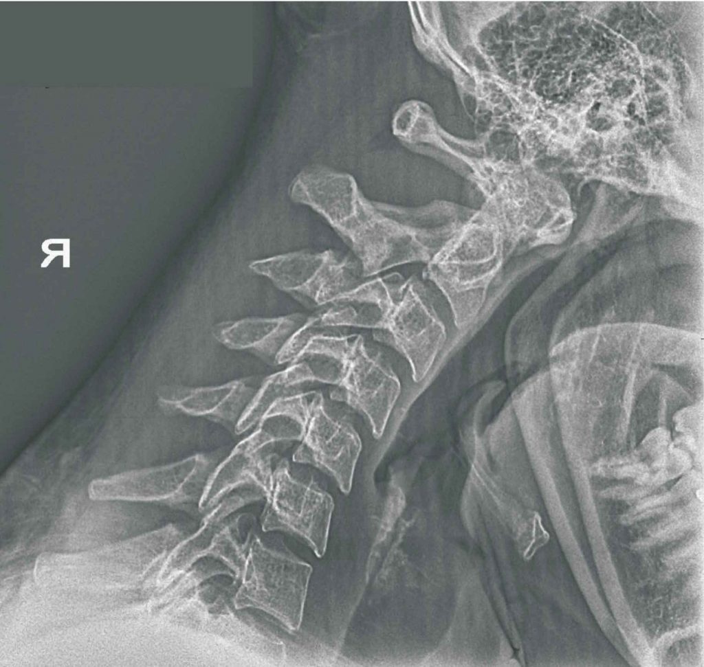
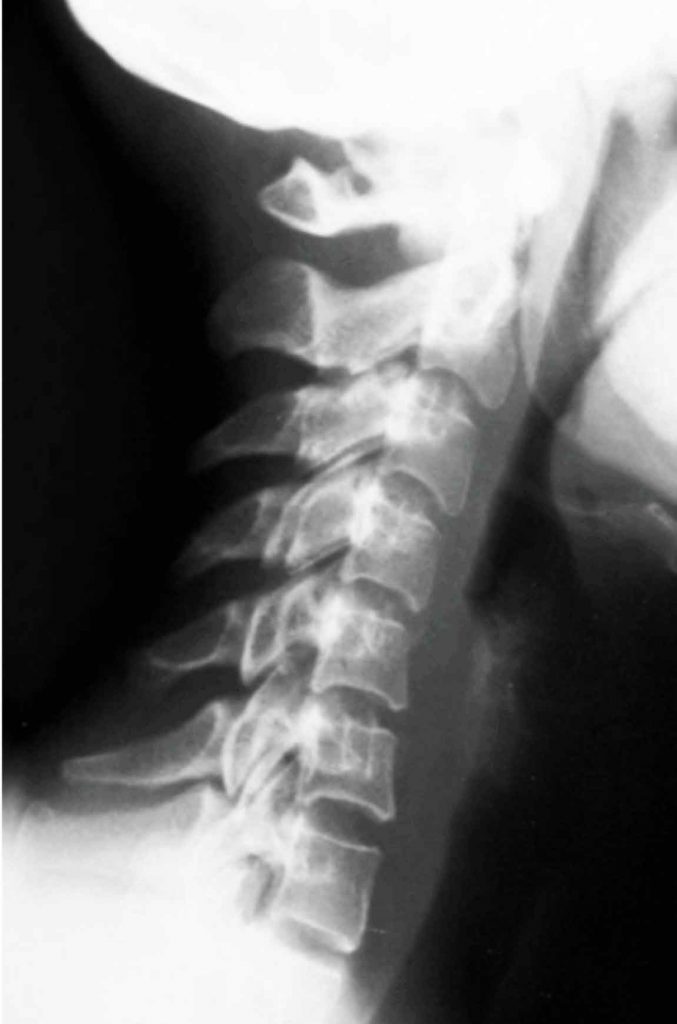
And finally, an important factor for my paradigm shift from film to digital – in fact, it was the essence of it – was the image itself. In the early days, whenever I looked at a digital film, whether of an 80-year-old or a 22-year-old, the skeleton appeared osteoporotic, which gave me the mistaken impression that digital was inferior. Was I ever wrong.
Actually, digital imaging more accurately depicts the proper and authentic trabecular pattern of the imaged bones. It provides more clarity to the final picture. This is a far truer picture of the skeleton we are examining. So why did I think that all digital films looked osteoporotic? Because the inherent limitations of the film/screen technology blurred the trabeculation artificially (read: artifactually), making all bones look somewhat whiter. Proof that plain-film x-ray, by its physical limitations, has historically and culturally forced us to accept, as normal, an inferior depiction of the reality of the skeleton being imaged. That’s not a bad thing; it was the accepted standard of the day, and that was the best we had – until digital came into the picture, literally.
We have better technology now than ever, and you and your patients deserve it. So that is my paradigm shift. I am very comfortable with it and I would not want to go back.
Dr. Marshall Deltoff, DC, DACBR, FCCR(C), is a specialist, clinician, professor, author and international lecturer. He has taken and read over 140,000 x-ray studies. He is the director of Images Radiology Consultants, where he offers complete online chiropractic x-ray reports to all doctors of chiropractic worldwide. Contact him at marshdel@yahoo.ca or visit his website: www.drmarshalldeltoff.com.
Print this page