Functional musculoskeletal assessment
By Anthony Lombardi
Features Clinical Techniques annex Anthony Lombardi assessment system Business talk chiropractors Chronic nociceptive stimuli dysfunction Functional musculoskeletal assessment musculoskeletal musculoskeletal assessment noxious stimuli Pelvic girdle Scapular girdle Vertebral columnHow assessment builds your practice
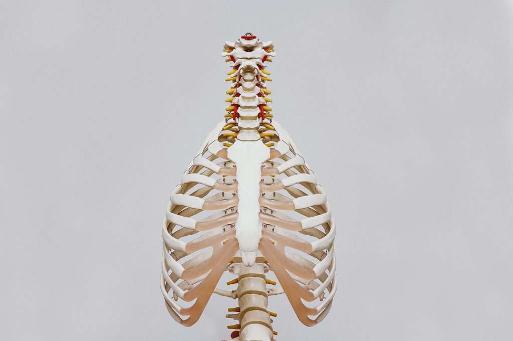 Photo by Nino Liverani on Unsplash
Too often, chiropractors label a patient’s condition in a way that does not provide specific information about the nature of their dysfunction. Terms like bursitis, tennis elbow, jumpers knee – even more general terms like low back pain, neck pain, and sciatica do little to define the nature of the injury.
Seven years ago I created (and started using) a functional musculoskeletal assessment system to help me assess, classify, and treat muscle- and joint-related injuries. In turn, my business practice exploded.
The key to this system and to clinical success, is locating muscle motor or neuromuscular inhibition in muscles that stabilize skeletal girdles (scapular/pelvic).
Muscles tend to “shut-off” or become inhibited for three reasons triggered by different forms of noxious stimuli:
- Trauma or repetitive strain injury (RSI)
- Pain
- Changes in the joint (arthritis/cartilage tears)
In the Clinical Journal Of Pain (2012),1 Nijs determined that nociception is most often processed without conscious thoughts. Therefore, in many cases, neither patients nor clinicians are aware of the interaction of the motor inhibition. Chronic nociceptive stimuli result in cortical delay of the motor output, thus creating reduced activity of the painful muscles. In addition, Nijs argues that nociception-induced motor inhibition might prevent effective motor retraining. This is of particular interest in my assessment approach because Nijs’ conclusion that motor inhibition might prevent effective motor retraining indirectly tells us that proper rehabilitation of our patient will not occur unless motor inhibition is revealed and corrected before actively retraining the impaired muscle.
Sign up to get the latest news and events from Canadian Chiropractor. Our E-newsletter will be sent to you only once per week, on Mondays.
By determining where the dysfunctions are, we can then make the appropriate treatment decisions to restore function and adaptability in our patients’ musculoskeletal systems.
My assessment approach focuses on the skeletal foundations of the musculoskeletal system which include:
- Vertebral column
- Scapular girdle
- Pelvic girdle
My goal is to assess the stability, strength and range of motion of the skull and extremities in relation to those foundations. Hamill (2006)2 described the girdles as foundations of human movement. This is because the shoulder and pelvic girdles protect and serve as adaptable attachment sites for muscles of the upper and lower extremities.
The skull is supported by muscles and soft tissue that anchor to the vertebral column (c-spine), while the scapular girdle anchors itself to the skull, vertebral column (c-spine) and humerus of the upper extremity. The vertebral column (t-spine/l-spine), which has soft tissue, attaches to the pelvic girdle that anchors to structures in the lower extremity beginning with the
femur.
Sedory et al. (2005)3 concluded that muscle groups in the proximal girdle of the kinetic chain of movement were associated with strength deficits in distal joint injuries. Bullock-Saxton et al. (1994)4 noted the influence of distal joint injury on muscle activation of proximal muscles of the pelvic girdle. This research was combined with contemporary ideas to formulate an assessment system that would encompass the tissues involved around the girdles, which are the centers kinetic movement.
Upper and lower body scans
A patient’s chief complaint determines which body scan(s) I perform to assess their range of motion, stability, and location of motor inhibition along the skeletal foundations.
For example, if the patient presents with anterior shoulder pain, I will perform the upper body scan. If the patient presents with left sided low back pain, the lower body scan will be performed. Typically, upper body musculoskeletal complaints will prompt you to perform upper body scans and lower body scans will be performed for lower body injuries.
The ultimate goal is to comfortably learn scans so that they can be completed in clinical practice within two to three minutes.
Here is a case study: A 42-year-old female presents with L lateral elbow pain of four months’ duration. She is a competitive tennis player and has had to modify her stroke to compensate for the elbow pain. NSAIDs and ice give her temporary relief.
My musculoskeletal assessment system revealed:
- On gait analysis that her right arm swings considerably less than her left
- The right trapezius is elevated during her gait
- The upper body scan reveals her cervical rotation (45 degrees on visual inspection) and lateral flexion is very limited to the right (10 degrees). Marked weakness occurs upon testing the supraspinatus in both full and empty can positions and her serratus anterior is inhibited.
Traditional Dx: R tennis elbow or lateral epicondylitis.
Traditional Rx: Treat local area with any number of different modalities and adjust the cervical and thoracic spines as needed.
Functional Dx: R lateral elbow pain associated with limited cervical spine movement and R scapulo-thoracic joint dysfunction.
Functional Rx: Restore motor inhibition in the scapular girdle and supraspinatus using a manual soft tissue technique like acupuncture or muscle release and then focus on restoring dysfunction in cervical/thoracic spines.
The results
What results from this assessment approach is that patients begin to see significant clinical changes in their conditions after their first, and/or second visit. This is dramatic for our patients and for our business practices, because patients will tend to tell everyone they know about the new chiropractor who solved their issues with a results-oriented approach. Remember, functional assessments are best suited for patients who have mechanical, sub-acute conditions. Chronic cases are typically more involved, and although functional assessment is a great start with these patients, it’s important to remember their condition is multi-faceted and so your approach will have to be as well.
Keep in mind that musculoskeletal assessment is more than just orthopedic tests – it’s an assessment of the adaptability of the mechanics of the body.
References
- Nijs, J et al. Nociception affects motor output: a review on sensory-motor interaction with focus on clinical implications. Clin J Pain. 2012; 28 (2):175-181.
- Hamill, J., Knutzen, KM. Biomechanical Basis of Human Movement. Lippincott Williams & Wilkins (2006).
- Sedory et al. Arthrogenic Muscle Response of the Quadriceps and Hamstrings With Chronic Ankle Instability. J Athl Train. 2005; 42 (3): 355-360.
- Bullock-Saxton JE, Janda V, Bullock, MI. The influence of ankle sprain injury on muscle activation during hip extension. Int J Sports Med. 1994; 15: 330-334.
Dr. Anthony Lombardi has presided over 103,000 patient visits in 14 years of practice at Hamilton Back Clinic (hamiltonbackclinic.com). Educational Materials are available at exstore.ca and acupuncturemotorpoints.com
Print this page