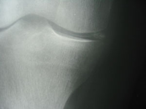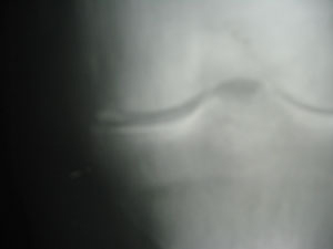
A 62-year-old woman presented with bilateral knee crepitus, swelling and stiffness.
Radiographic examination reveals bilateral chondrocalcinosis. (Figures A and B)
Diagnosis: Calcium pyrophosphate dihydrate (CPPD) deposition disease; also known as pseudogout.
Advertisement
 |
 |
| Figure A | Figure B |
Some points to remember about CPPD:
- inflammatory joint disease resulting from deposition of calcium pyrophosphate dihydrate into the synovial fluid; a metabolic disturbance in which there is an impaired degradation or excessive production of pyrophosphate
- may be acute (more prevalent in males) or chronic (more prevalent in females)
- frequently associated with diabetes mellitus
- features include articular cartilage calcification, with possible periarticular calcification of synovium, articular capsule, tendons, bursae
- affected hyaline cartilage appears as a fine radiopaque line parallel to the contour of the articular cortex
- affected fibrocartilage demonstrates a characteristic punctate calcification
- in the knee, the menisci classically demonstrate calcification
- joint destruction associated with CPPD is virtually indistinguishable from that of osteoarthritis
- CPPD also tends to affect the shoulder, elbow, radiocarpal joint, hip, symphysis pubis and patellofemoral joint
- joint aspiration may be necessary to confirm the diagnosis •
Print this page
Advertisement
Stories continue below
Related