
Magnetic resonance imaging (MRI) is a non-invasive imaging technique
that does not involve exposure to ionizing radiation. MRI uses a
powerful magnetic field, radiofrequency pulses and a computer to
generate high-quality images of any part of the body.
The author would like to thank Paul Ouimet, DC (HealthQuest Chiropractic) for his involvement in the clinic and assistance in writing this article.
Magnetic resonance imaging (MRI) is a non-invasive imaging technique that does not involve exposure to ionizing radiation. MRI uses a powerful magnetic field, radiofrequency pulses and a computer to generate high-quality images of any part of the body. The anatomic detail of soft tissue structures such as cervical spine ligaments, pelvic floor muscles, brain and spinal cord are all seen in great detail with MRI. It can demonstrate structural abnormalities, injuries, tumours, infections and other disease processes in all areas of the body better than any other imaging method.
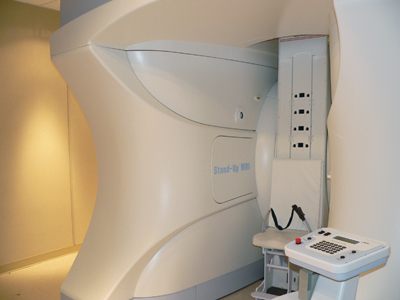 |
| The Fonar upright/dynamic MRI unit. Photo by Michael Koehn, reprinted with permission |
Most MRI machines consist of a narrow horizontal tube with a table that moves into the bore of the magnet. Images are obtained with the patient lying flat on the table. The procedure is lengthy, cramped, noisy and is often not tolerated by claustrophobic or large patients. As well, it does not enable the patient to be imaged in the position that generates his/her symptons.
The quality of an MRI unit is generally judged on the basis of the field strength of the magnet. The higher the magnetic field (in Tesla) the better the image quality. Most machines in clinical use are 1.0 or 1.5 Tesla units. 3.0 and 7.0 Tesla units are used mostly for research. However, field strength is not the only variable that determines the image quality. Image acquisition times, coil configuration and a number of other variables can improve image quality in lower field strength magnets. Lower field strength magnets produce less artifact from implanted metals such as screws, rods and plates and can produce images superior to higher field strength magnets in patients who have had prior surgery with metal implants.
NEW TECHNOLOGY IN MRI
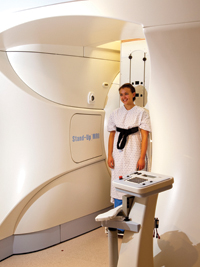
Dynamic MRI refers to the ability to acquire images with the patient in positions other than lying flat on their backs. This has the advantage of acquiring more information about the anatomy and pathology in weight-bearing and bending postures such as sitting, standing, flexing forward or backward or lying down.
 |
|
| A patient undergoing imaging in the upright MRI unit Photo by Michael Koehn, reprinted with permission |
Back and neck pain are among the leading complaints for which patients seek treatment from chiropractors, physiotherapists, massage therapists and physicians. These patients often have pain when sitting, standing or bending and get relief from their pain by lying down. Consequently, the recumbent MRI is unable to achieve a complete and accurate diagnosis of the patient’s problem.
Pressure measurements inside lumbar discs have shown the pressure to be lowest when the patient is lying down and as much as 12 times higher when the patient is sitting and bending forward.1 Therefore, taking an MRI with the patient in a supine position may not adequately reveal the cause of the patient’s pain in a significant percentage of cases.
IMPROVED DIAGNOSIS WITH DYNAMIC MRI
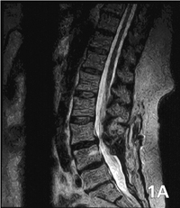
MRIs performed with patients sitting with flexion and extension views have uncovered “hidden” abnormalities such as disc herniations, spinal stenosis and spondylolisthesis in patients who had a “normal” recumbent MRI. One study of 510 patients reported an 18 per cent incidence of spondylolisthesis of greater than three millimetres that were missed in neutral MRIs, but that were found on flexion and extension MRIs.2
|
|
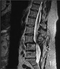
| The recumbent scan (1A) is from a patient with recurrent low back pain following L4-5 fusion. Note the multilevel bilateral laminectomy. The upright scan (1B) shows an anterior slip of L3 on L4 associated with stenosis of the central spinal canal at L3/4 at the level above the surgical spinal fusion. The hypermobile intersegmental instability is visible only when the patient is scanned upright. Case and images courtesy of M.S. Rose, MD | |
 |
|
 |
|
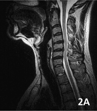
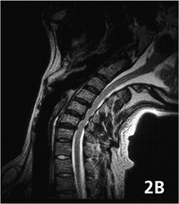
| The upright extension image (2B) demonstrates marked stenosis of the central spinal canal and compression of the underlying spinal cord resulting from further posterior disc protrusions extending into the anterior spinal canal and focal ligamentous unfolding posteriorly. This compression of the underlying spinal cord is not evident on the recumbent scan. (2A) Case and images courtesy of Melville MRI | |
 |
|
|
|
Flexion and extension views of the cervical and lumbar spine can demonstrate instability, changes in the size and shape of disc herniations, nerve root compression, changes in the degree of spinal stenosis, ligament injuries and facet joint abnormalities much better than recumbent imaging. Positional MRI reportedly shows nerve root compression in 24 per cent of patients who had no nerve root compression identified on conventional recumbent MRIs.3 The degree of disc bulge and herniation increased in 19 per cent of patients in seated flexion and extension MRI scans compared to upright neutral MRI scans.4
Failed back surgery refers to patients who do not get pain relief following a back operation. This is probably due to obtaining the wrong diagnosis rather than performing a bad surgical procedure. Positional MRI could improve diagnostic accuracy and lead to more effective treatment protocols and fewer failed back operations.
Symptoms from Chiari I malformations are often positional and the full extent of cerebellar tonsil herniation is demonstrated better on MRIs with the patient seated rather than supine. The ability to image joints such as ankles, knees and hips with patients’ weight-bearing could provide additional information about ligament and meniscal abnormalities better than those seen in the recumbent position. Scoliosis can be imaged by MRI in the upright position far better than with X-rays. Upright MRI provides a view of not only the vertebrae, but also the soft tissue and the angles of rotation of the vertebrae. There are many situations in which more information could be obtained from MRI scans with patients in positions other than lying flat on their backs during the study. Some of these benefits have been demonstrated, while other possible advantages to this technology are still awaiting clinical studies to prove the benefits of dynamic MRI imaging. As well, there are many research opportunities for dynamic MRI imaging that will increase our understanding of disease and improve treatment.
Another important point to note is that an MRI exam, unlike X-ray, is radiation-free. The National Cancer Institute reported a 70 per cent increase in breast cancer in women with scoliosis who had repeated chest x-ray examinations throughout their childhood and adult life.5
OPEN CONFIGURATION, UPRIGHT, MULTIPOSITION MRI
Fonar Corporation (www.fonar.com) is a leading developer of MRI technology and, currently, the only manufacturer of the upright MRI.
The company has produced a 0.6 Tesla upright, multiposition MRI with a vertical bore and an open configuration. It can acquire images of any body part and in any plane with the patient sitting, lying, standing and bending.
The upright MRI allows patients to sit and watch television in a quiet, comfortable position while they have their MRI. Fonar has also developed an assortment of gradient coils that enhance image quality at the lower field strength. For imaging of the brain, spine and many joints the image quality is equal to those obtained in a 1.5 tesla magnet.
There are approximately 150 upright MRI scanners worldwide. There is currently only one in Canada, located in Kamloops, British Columbia.
DYNAMIC MRI – IMPLICATIONS FOR CHIROPRACTIC
Spinal adjustments have been practised for thousands of years and are the basic foundation for chiropractic care. The idea of the “spinal subluxation complex” has been used by chiropractors, but can be difficult for physicians to envision or accept. Dynamic MRI could become integral to chiropractic practice, particularly with respect to enhancing diagnostic acumen, as well as in chiropractic education and research. Performing positional MRIs before and after spinal adjustments could identify the anatomic or pathological correlation between spinal dysfunction and pain. This technology could also validate current treatment protocols and aid in the development of new chiropractic procedures. •
REFERENCES:
- Nachemson AL. 1981 Disc pressure measurements. Spine 6:93-97.
- Hong SW et al. 2007. Missed spondylolisthesis in static MRIs but found in Dynamic MRIs in patients with low back pain. The Spine Journal 7 (5S):69S.
- Weishaupt D et al. 2000. Positional MRI imaging of the lumbar spine: does it demonstrate nerve root compromise not visible at conventional MR imaging? Radiology 215 (1):247-253.
- Zou J et al. 2008. Missed lumbar disc herniations diagnosed with kinetic magnetic resonance imaging. Spine 33 (5):E140-E144.
- National Cancer Institute. 2000. http://www.cancer.gov/newscenter/scoliosis2000.
Dr. Richard Brownlee received BSc (University of Western Ontario), MSc (University of Toronto) and MD (University of Calgary) degrees prior to completing his neurosurgery training at the University of Calgary in 1996. He has had an active cranial and spinal practice in Kamloops, British Columbia, and is president of the new Welcome Back MRI and Pain Management Centre in Kamloops, British Columbia, which is the site of the only upright MRI unit in Canada. (www.welcomebackclinic.com).
Print this page