
Rasterstereography: Radiation-free technology for spine and pelvis analysis
By Jean-Pierre Gibeault PEng
Features Clinical Techniques 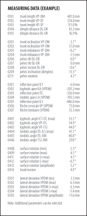 |
| Fig. 1 |
After more than 20 years of research in Europe, the technology of body surface measurement by rasterstereography, combined with biomechanical modelling techniques, has produced a device for the fast, radiation-free evaluation and analysis of the spine and pelvis. This German device is used by universities and research clinics. It has also been utilized in, primarily private, chiropractic, orthopedic and dental clinics for diagnostics and for specific and interdisciplinary therapies. With the first units produced for commercial use in 1996, this technology is now in its fourth generation. Four hundred and ninety units have been produced commercially and most of these are used in Germany. The high accuracy of this technology, used on scoliosis patients, was clinically confirmed by Drerup et al (1997) and compared with customary thorax radiographs by Hackenberg et al (2003).
Origin and scientific foundation of this technology
The aspects of spinal deformity that have been researched extensively at many universities and research centres are the shape of the back, shape analysis of the body surface and techniques of body surface measurements.
The objective driving this research in its early days – around 1980 – was to develop technologies and devices that would be complementary to radiology for the evaluation and monitoring of patients suffering from idiopathic scoliosis. The need for frequent follow-up evaluations during therapy, and the necessity to greatly reduce the overall radiation load during the duration of the therapy, were strong motivators to develop accurate and reliable devices.
 |
| Fig. 2 |
Among many research centres, the Institute for Experimental Biomechanics and the University Orthopedic Clinic at the University of Münster in Germany continuously develop a large body of research in this area. Research papers have been – and still are – produced by many of their professors, researchers and doctoral students.
An optical system for spinal and pelvis analysis
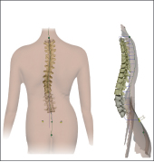
One of the ways to meet the objective of developing a radiation-free, fast, contactless and reliable device to complement X-ray measurement systems is to use the combination of 3-D-shape measurements and biomechanical modelling to reconstruct and display the spine structure and calculate the key spinal and pelvic parameters. (Figures 1 and 2)
Capturing and measuring the dorsal profile (back shape)
 |
| Fig. 3 |
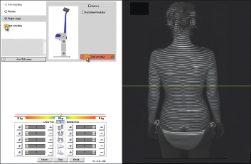
White Light Raster Line Triangulation (WLRT) enables the scanning of objects in 3-D by projecting raster lines on their surfaces and by capturing these lines under a known and fixed angle with a camera. (Figure 3) Based on triangulation algorithms, spatial co-ordinates of all raster points are calculated, resulting in a dense point cloud of randomly distributed points describing the measured surface. These data points are transformed to a regular grid by using interpolation, which will simplify further calculations. In this way the system captures and analyzes a body shape, statically or dynamically.
 |
| Fig. 4A |
Rasterstereography excels by its high precision – error margin ≤ 0.1mm – and allows a radiation-free representation of the profile. The speed of the measurement is fast at 0.04 seconds and the total dorsal surface is registered simultaneously.
The recognition of the anatomical structure through the automatic identification of anatomical landmarks on the body surface provides the basis for a reconstruction of the 3-D profile of the dorsal surface.
Mathematical construction and display of the spine structure
The aim of capturing, measuring and analyzing the dorsal surface – back shape – is to obtain information about the 3-D shape of the vertebral column.
It is well documented that the vertebral rotation is correlated to the surface rotation and this allows us to establish the relationship between the back shape and the shape of the spinal midline. In our case, the surface rotation is measured by the angle of the surface normal, or horizontal component. To do this, the high sampling density and resolution provided by rasterstereography is essential.
 |
| Fig. 4B |
The calculated construction of the spinal midline requires three inputs:
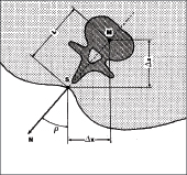
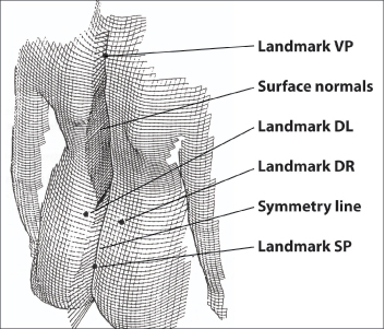
1. The line of the spinous process.The line of the spinous processes is estimated by the symmetry line of the back. The symmetry line (solid line in Figure 4A) is composed of the symmetry points of the horizontal profiles. A symmetry point, in turn, is defined by that point which divides the profile into two halves with minimum lateral symmetry (with respect to surface curvature). For the model, we assume the symmetry line to be a representation of the line of the spinous processes and it is a generalization of the medial sagittal profile.
2. Surface rotation. As mentioned above, we measure surface rotation by the horizontal component of the direction of the surface normal. From any grid point in Figure 4A, components of the surface normal are known from curvature analysis. On the symmetry line these values are calculated by interpolation. In Figure 4B, the surface normals are represented by bars erected on the symmetry line. As the results show, it is reasonable to assume that the horizontal component of the normal angle is equal to vertebral rotation.
3. Anatomical landmarks. An automatic recognition of four anatomical landmarks (vertebra prominens (VP), sacrum point (SP), right crista iliaca posterior superior (DR), and left crista iliaca posterior superior (DL) by means of the connected software provides the basis for a reconstruction of the three-dimensional profile of the dorsal surface.
 |
| Fig. 5 |
The landmarks are used for skeletal reference of the surface data. In particular, the vertebra prominens landmark is used as the origin of a body-fixed co-ordinate system both for radiography and for surface measurements. Furthermore, trunk length, trunk imbalance, pelvis inclination, and similar parameters may be determined from these landmarks. In Figure 4A the landmarks are represented by black dots.
Curvature analysis is combined with an algorithm for data smoothing and calculation of the surface normals. As a byproduct, the original measurement points are transformed into a regular square grid over the frontal (x-y) plane. The result of this procedure is presented in Figure 4B.
What does rasterstereography offer the clinician?
 |
| Fig. 6 |
Using a fast – 40-millisecond – high-definition optical measurement of the surface of the back of your patient, rasterstereography produces graphical, clinical and analytical information about the spine, the pelvis and the posture.
It takes only 40 milliseconds to do a scan and the results are available immediately on your screen and printed on the spot.
Ideal for chiropractors, the rasterstereography provides clinical information to enhance your diagnostics and develop treatments, and document your treatment outcomes.
Rasterstereography is contactless, radiation-free and non-intrusive and allows chiropractors to perform multiple scans of each patient – e.g., before and immediately after an adjustment – and a typical report will offer the capability to compare four sets of results taken at different dates.
A complement to your radiology equipment, rasterstereography does not require any licence or special building construction permit, nor any lengthy training for its accurate and repeatable operation.
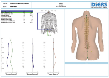
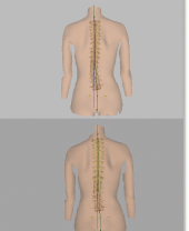
A typical report is shown in Figure 5. This system is an ideal tool for chiropractors, as it allows multiple radiation-free scans tha contain key information on the spine and pelvis complex of each patient. With rasterstereography, chiropractors can first perform a detailed evaluation, to help determine a treatment plan, and then monitor and document, the progress of this treatment plan. This is demonstrated in Figure 6. Patients can receive updates of the progress of their treatment and can also easily be handed a copy of their scans for their own records.
Various applications for rasterstereo-graphy technology have been developed and are currently being investigated in clinical situations. In the months to come, results from these investigations will be collated and available to chiropractors who may be interested in incorporating this device into their diagnostic regimes.
(Canadian Chiropractor is pleased to have been offered the opportunity to update our readers with results from clinical trials with this technology – which is new to Canada – in another issue later in 2008. Please look for more on rasterstereography in our September issue.)
References:
Drerup B, Hierholzer E, Ellger B. Shape analysis of the lateral and frontal projection of spine curves assessed from rasterstereographs. In: Sevastik JA, Diab KM, eds. Research Into Spinal Deformities. 1st ed. Amsterdam, The Netherlands: lOS Press; 1997:271-275.
Hackenberg L, Hierholzer E, Potzl W, Götze G, Liljenqvist U. Rasterstereographic back shape analysis in idiopathic scoliosis after posterior correction and
fusion. Clin Biomech.2003;18:883-889.
Hackenberg L, Hierholzer E, Potzl W, Götze C, Liljenqvist U. Rasterstereographic back shape analysis in idiopathic scoliosis after anterior correction and fusion. Clin Biomech. 2003;18:1-8.
Turner-Smith AR, Harris JD, Houghton GR, Jefferson RJ. A method for analysis of back shape in scoliosis. J Biomech. 1988;21(6):497-509.
Jean-Pierre Gibeault, PEng, is the founder and CEO of Biometrix
Medica. He graduated from Queen’s University with a degree in
electrical engineering and has been involved in scientific
instrumentation throughout his career. Headquartered in Canada, his
companies deliver medical products and equipment in the Americas and
Scandinavia.
Print this page