The Oolo-Austin Test – also simply
known as “the Austin Test” – is an orthopedic examination used to
assess the axial rotation range of motion of the thoracolumbar spine.
 |
| Diagram 1 |
The Oolo-Austin Test – also simply known as “the Austin Test” – is an orthopedic examination used to assess the axial rotation range of motion of the thoracolumbar spine. The test is named after the founder of Trigenics“ Myoneural Medicine, Dr. Allan Oolo- Austin. The test is a range of motion assessment used in conjunction with the Trigenics® neuromanual treatment system. It primarily targets the multifidus and erector spinae muscles of the back.
Muscles are the source and the recipient of the greatest amount of neural activity in the body(1). Trigenics is based on detecting and correcting sensorimotor dysfunction causing neuromusculoskeletal and articular conditions. It is the philosophy of the Trigenics practitioner that the etiology of such conditions lay first in the aberrant sensorimotor control of the holding and moving elements, which traverse the associated joints(1) controlling or restricting movement. The secondary causes in chronic cases are often due to resultant dysfunction of associated and related tissues, which form a “neuropathophysiologic dysfunctional kinetic chain continuum”(1). By using the Oolo-Austin Test one is able to determine which muscles of the spine are in a hypertonic, overfacilitated state or conversely those that are in a hypotonic, inhibited state.
Numerous studies have been performed that link dysfunction of the multifidus and erector spinae spasm with chronic low back pain and loss of range of motion(7). Notables such as the late Vladimir Janda, who did extensive studies on functional neurokinetic imbalance patterns due to dysafferentation(1,7). Janda was a neurologist who later specialized in manual medicine and rehabilitation. His extensive understanding of both practices allowed him to explain chronic pain syndromes in a manner that integrated neurologically based principles with manual medicine procedures and techniques(6). Chiropractic and osteopathic manipulative procedures has been proven effective in the restoration of range of motion in chronic back pain sufferers; however, osseous manipulation procedures have not always proven to be enough, as this does not correct the root cause of the problem, which is “aberrant post-trauma neurology”(1). This is where the Trigenics myoneural treatment system plays an intricate role in the correction of aberrant neuromotor control.
In developing his test, Oolo-Austin points out that a study by Kuriyama et al. measured the electromyography activity of the multifidi muscles and the longissimus muscles during active lumbar range of motion(4). The study utilized Kinesiologic electromyography (EMG) to record muscle activity, in standing resting position, and also during active trunk forward flexion and extension, lateral bending and axial rotation. No muscular activity was observed in the full trunk flexion position in the control group, whereas continuous muscle activity was observed in the low back pain group(4). On axial rotation, an intermuscular time lag was observed at the beginning of the motion in the control group, however, in the low back pain group, there was no such time lag(4). Kuriyama demonstrated that the paraspinal muscle activity restricted lumbar range of motion to protect from injury by movement. It was found that reduced intervertebral range of motion has also been seen previously in patients with low back pain with degenerative changes in the lumbar spine (4). Kuriyama concluded that restricted intervertebral motion in the patients may have been due to continuous intrinsic muscle activity, working to protect from injury of the joint, intervertebral disc and ligament(4). The Oolo-Austin test is used to identify those particular muscles of the spine that in are in a neurologically over-facilitated or protective state. A large part of the Trigenics Myoneural Assessment involves muscle length testing for overfacilitation. (Conversely strength testing is also used to measure neural
inhibition) The Oolo-Austin test is strictly used to assess the length of the surrounding musculature of the spine, in particular the multifidus and the erector spinae.
 |
| Diagram 2 |
THE OOLO-AUSTIN TEST PROCEDURE
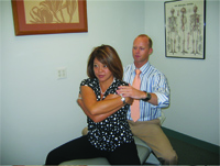
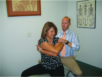
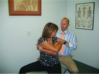
The practitioner sits directly behind the patient who is in a seated position, straddling the treatment table with their arms crossed over their chest (diagram 1). The practitioner assesses to see if the patient has equal range of axial rotation of the head and neck and then passively rotates the patient by contacting the patient’s shoulders to pull with one hand and push with the other. The patient is passively rotated until they reach end range of motion (diagram 2) and the patient is asked to look back at the practitioner. The rotational component can be measured either visually by the practitioner viewing the amount that can be seen of the eye of the patient farthest from him or by using a goniometer for a more accurate reading. By having the patient look into the direction of rotation when they have reached the passive end range, they are able to actively take part in the pre- and post-treatment assessment and objective findings (diagram 3). The doctor will often note that, in one direction, both eyes of the patient can be seen but in the opposite direction, only one can be seen, indicating a difference in rotational ability. A unique aspect of the Trigenics treatment system is that it encourages the patient to take an active role in their treatment. The test is done bilaterally, and may need to be performed more than once in order to correctly determine the side of reduced axial rotational range of motion. A decreased axial rotation range of motion will be evident when the surrounding stabilization muscles of the spine are in a hypertonic state. Any difference in axial rotational range of motion from one side to the other would constitute a positive Oolo-Austin test.
In order to fully understand the purpose of the Oolo-Austin test, we must first have a good understanding of the muscles that are being tested. According to Hansen et al., “In order to fully determine the segmental actions of a muscle, it is necessary to know the exact sites of attachment as well as the lines of action with respect to the joints it crosses”(2).
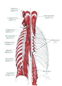
The back has multiple layers of muscles. The more superficial muscles of the back are considered the prime movers and include the trapezius, latissimus dorsi, rhomboid major, rhomboid minor, and levator scapulae. The intermediate layer consists of the serratus posterior superior and the serratus posterior inferior(5). For the purpose of this article, I am going to focus on the deep muscles of the back that act directly on the vertebral column. Both the erector spinae and the multifidus are crucial muscles for movement and stabilization of the spine respectively(5).
The lumbar and thoracic erector spinae muscles include the iliocostalis, longissimus, and spinalis. Upon unilateral contraction, the erector spinae laterally flex and ipsilaterally rotate the spine, and upon bilateral contraction, they extend the spine and depress the ribs(2). According to Gray et al, the erector spinae originate from the anterior surface of a broad and thick tendon, the erector spinae aponeurosis(2). The muscle fibres form a large fleshy mass, which in the upper lumbar region, splits into three columns: spinalis (medial), longissimus (intermediate), and iliocostalis (lateral). The muscles lie in the groove on the side of the vertebral column, lateral to the multifidi, and are covered by the thoracolumbar fascia. In general, the erector spinae cross the lumbar region without attachment to the lumbar vertebrae(2).
The musculotendinous fibres of the lumbar erector spinae consist of two parts – a medial and a lateral division – which are labelled as longissimus thoracis pars lumborum and iliocostalis lumborum pars lumborum, or simply a superficial and deep part(2). Each fascicle attaches directly to the iliac crest. The fascicle of both parts lumborum (longissimus and iliocostalis) can be resolved into horizontal and vertical vectors(2). Unilateral contractions flex the vertebral column in a lateral direction, and bilateral contraction produces posterior sagittal rotation (2). Thus, they are well suited to co-operate with multifidi to oppose the flexion effect of the abdominal muscles when they act to rotate the trunk(2).
 |
| Diagram 3 |
The deep paraspinal muscles include the semispinalis, multifidi, and the deep rotators(2). Upon unilateral contraction, they laterally flex and contralaterally rotate the spine, and upon bilateral contraction, they extend the spine(2). The multifidi consist of a number of fleshy and tendinous fasciculi, which lie deep to the foregoing muscles and fill the groove at the sides of the spinous processes of the vertebrae, from the sacrum to the axis (C2)(2). According to Gray et al., they are capable of producing extension, lateral flexion, and rotation(2). However, the study by Hansen et al. shows that when the multifidi are studied only as individual muscles, they seem to act more as stabilizers rather than prime movers of the vertebral column(2). They used EMG to confirm this result, in which it was found that the multifidi play a role in controlling intersegmental motion(2).
The rotational components of these two muscle groups, work antagonistically to each other whereas the opposite sides of each group work synergistically. When considering the spine as a whole, it consists of multiple vertebrae with a wealth of possible movements(2). However, it is the tissues connecting the vertebrae as well as the shape and orientation of the facets that constrain the movement considerably. Thus, the spine relies on its ability to constrain movement between the vertebrae to patterns also known as coupled motions, which are compatible with the limitations on tissue(2).
Upon passive axial rotation of the lumbar spine, the ipsilateral multifidi and the contralateral erector spinae are lengthened. When you passively rotate the patient to the right, their right multifidi and left erector spinae muscles would be lengthened and their left multifidi and right erector spinae would be shortened. For example, when performing the Austin test on a patient, you notice that they are not rotating to the left as far as they are to the right. A patient that presents with less axial rotational range of motion to the left would indicate that either the left multifidi and/or the right erector spinae are in a hypertonic or neurologically facilitated state. Another presentation may be if the patient has normal axial rotational range of motion to the right and excessive axial rotational range of motion to the left. This would indicate that the patient would need a strengthening procedure to increase tonicity of the left multifidi and right erector spinae. The Oolo-Austin test is a quick and efficient method that allows the practitioner to accurately assess the deep back muscles resetting of neuromotor gamma gain in order to be lengthened and those which need to be facilitated and thereby strengthened. A physician or therapist practising manual medicine who has trained to become a registered Trigenist Physician/Practitioner uses either a Trigenics Strengthening procedure, referred to as “TS” or a Trigenics Lengthening procedure referred to as “TL.” This is carried out in order to correct the aberrant neuromotor control, which causes an imbalance in the pulling forces on the joint with consequent dysfunctional athrokinetics.
A study was performed by Richardson et al. aimed to examine EMG activity of abdominal and back muscles as well as triaxial torque output during isometric axial rotation at different exertion levels in back pain patients and matched controls(3). The findings of the study demonstrated that decreased activation of one muscle may be compensated by over-activity in other muscles(3). The examiners deduced from this finding that spinal stability could be compromised due to the reduced levels of activity of the multifidi muscles during axial rotation exertion in back pain patients(3).
 Discussion of Treatment
A patient who presents with a positive Oolo-Austin Test would likely benefit greatly from a combination of joint manipulation, to correct any aberrant osteology and arthrokinetics, and soft tissue techniques to treat aberrant histology. However, the imbalanced biomechanics with resultant pain and dysfunction will continue to reoccur unless the root cause of the problem is addressed, that being aberrant neurology, also known as dysafferentation. It is for this reason that Trigenics may be being the “missing element” to total sustainable care(1). It is important to note that Trigenics is not a technique, and its primary objective is not adhesion removal nor the mechanical treatment of soft tissues(1). Trigenics is actually a “global neurological treatment system” with the primary objective to correct sensorimotor malfunction(1). It incorporates applying three treatment “techniques” simultaneously for an amplitude summation effect neurologically; “Trigenics combines functional reflex neurology with mechanoreceptor manipulation and cerberopulmonary biofeedback”(1). The objective of this treatment system is to change the “tone” or “gamma gain” within the tissues by altering the afferent and efferent neural signals(1). Dr. Oolo-Austin and many of his students have successfully reported treating hundreds of patients using Trigenics. Numerous physicians and therapists have found it remarkably effective in providing correction, functional restoration and/or relief for a wide range of neuromusculoskeletal conditions and chronic pain syndromes.(1).
The Oolo-Austin Test is an efficient method of assessing a patient’s thoracolumbar axial rotation and range of motion with the purpose of detecting aberrant intrinsic neuromotor control and function. The Trigenics treatment system, when applied to dysfunctional multifidi and erector spinae has been found clinically to be very effective in alleviating low back pain as well as increasing axial rotation as well as trunk flexion. Often patients complaining of severe debilitating back pain with inability to fully forward flex will respond dramatically well with significant decrease in pain and increase in ROM post treatment using the Trigenics® Applied-Functional-Neurology®
procedures.
References:
1. Oolo-Austin A. DC. Trigenics Myoneural Medicine – Theory Manual. Toronto, The International Institute of Trigenics, Inc. 2005.
2. Hansen L, PhD, de Zee M, PhD, Rasmussen J, PhD, Andersen TB, PhD, Wong C, PhD, Simonsen EB, PhD, “Anatomy and Biomechanics of the Back Muscles in the Lumbar Spine With Reference to Biomechanical Modeling.” Spine. 2006; 31(17):1888-1899.
3. Ng JK, Richardson CA, Parnianpour M, Kippers V, EMG activity of trunk muscles and torque output during isometric axial rotation exertion: a comparison between back pain patients and matched controls.
J Orthop Res. 2002; 20(1):112-21.
4. Kuriyama N, Ito H, Electromyographic Functional Analysis of the Lumbar Spinal Muscles with Low Back Pain. J Nippon Med Sch. 2005; 72: 165-173.
5. “The Anatomy Lesson.” <http://mywebpages.comcast.net/wnor/homepage.htm>. 1999 by Wesley Norman, PhD, DSc. Muscles of the Upper Limb and Back. June 20. 2007. <http://mywebpages.comcast.net/wnor/lesson6musclesofback.htm>
6. Craig Morris, DC, DACRB, FAFICC, CSCS. “The Enduring Message of Vladimir Janda, MD, DSc”, Dynamic Chiropractic, April 21, 2003, Volume 21, Issue 09, <http://www.chiroweb.com/archives/21/09/19.html>
7. Frank M. Painter, DC. Updated 8-24-2006 “Vladimir Janda Citation List.” June 20, 2007. <http://www.chiro.org/LINKS/
ABSTRACTS/Janda.shtml>
Print this page