
In our last edition of Technique Toolbox, we began with a sample case
about a 40-year-old carpenter who presented with headaches and low back
pain and on whom leg length analysis was performed resulting in
ipsilateral contracture upon turning the head to the right and left.
In our last edition of Technique Toolbox, we began with a sample case about a 40-year-old carpenter who presented with headaches and low back pain and on whom leg length analysis was performed resulting in ipsilateral contracture upon turning the head to the right and left.
 |
|
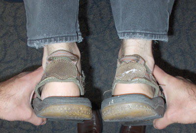
| Photo 1: Patient presents with a contracted right leg in the extended position. |
|
 |
|
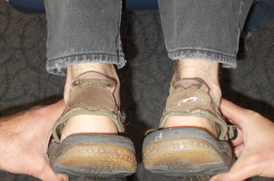
| Photo 2: The right leg continues to pull short in the flexed position, indicating a possible Derefield Negative. Advertisement
|
We mentioned that this case was dealing with a category called a Bilateral Cervical Syndrome (BCS) – wherein the occiput has subluxated anterior-superior – and reviewed a classic Thompson Technique prone adjustment to correct this subluxation pattern. Following the BCS adjustment, the doctor rechecked the patient’s leg lengths and noticed that the patient no longer presented with even legs but demonstrated a contracted right leg in the extended position. Furthermore, when the doctor lifted the patient’s legs to 90 degrees, he noticed that right leg continued to pull short.
What does this new leg length analysis indicate? Did the doctor do something wrong with the occiput adjustment? Are there other subluxations that now need to be addressed and corrected? I will answer these questions and more in this issue of Technique Toolbox, as I review the Thompson Technique’s Derefield Negative (D-).
History
I have summarized the history of the Thompson Technique in Part 1 of this article, and I would now like to focus on the fact that, paramount to the Thompson Technique is the use of leg length analysis, which is originally credited to Dr. Romer Derefield. Leg length analysis provided a consistent reference tool to be utilized throughout the adjusting procedure. Again, due to the science limitations of his era, Clay could not scientifically explain why the technique worked. But new neurological and biomechanical information can now be harnessed and I have been able to establish a more scientifically based understanding of the mechanisms underlying the technique.
So, what has happened in our original case? Did the doctor do something wrong with the original adjustment, and what exactly is a D-?
Once the occiput was corrected, the leg pulled short in the extended position and stayed short in the flexed position. The answer to the above questioning is, the doctor didn’t do anything incorrectly with the occiput adjustment – this change in leg length findings simply indicates that further subluxations exist, in addition to the occiput subluxation.
So, what should the doctor proceed to do now?
Step 1: Analysis
(See Photos 1-2)
After clearing the occiput, a short leg presents that is different from the pre-adjustment situation. The doctor must now rule out if any cervical subluxations exist by having the patient again rotate their head to the right and left. In this case, head rotation did not balance the legs in the extended position, indicating that the cervical spine is clear. If the cervical spine were subluxated, head rotation to one or both sides would have balanced the legs in the extended position. Since that did not occur, the doctor will move on to another primary subluxation area – the pelvis.
In this case, the right leg appeared short in the extended position, and remained short in the flexed position – this could indicate that a possible D- exists, provided that one of the following tender points is elicited:
- ipsilateral medial-proximal tibia
- ipsilateral ischial tuberosity
- ipsilateral PSIS
- ipsilateral pubic bone
- contralateral erector spinae from T2-T6
Note that ipsilateral and contralateral are referenced in relation to the short leg.
Please remember that only one of the aforementioned tender points needs to be elicited, not all of them. If one tender point is present, this verifies that the problem is a D-, which indicates that the sacral base has subluxated anterior-inferior (AI) on the ipsilateral short leg side.
Step 2: Correction – Modified Sacral Prone Adjustment
(See Photos 3-5)
|
|
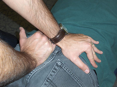
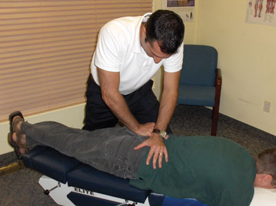
| Photo 3: D- contacts displayed. Note how the pisiform is on the contralateral sacral apex, and the stabilization hand contacts the ipsilateral PSIS. |
Photo 4: Note how the affected side leg crosses over the unaffected side, opening up space in the affected SI Joint. |
 |
|
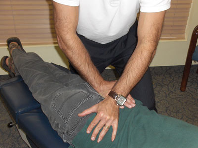
| Photo 5: Doctor and patient positioning is displayed. Note how the doctor is standing on the contralateral side of the subluxation. |
There are several ways of correcting this subluxation. The following is the Modified Prone Sacral Adjustment:
- Patient: Prone. Cross the short leg over the opposite leg to gap the involved joint.
- Doctor: Opposite side of the lesion.
- Table: Pelvic piece in the ready position.
- Contact: Fleshy hypothenar on opposite sacral apex.
- Stabilization: PSIS of short leg side, or reinforcing the contact hand.
- LOC: P-A with torque towards involved side. Repeat three times.
It is important for the doctor to note that crossing the affected leg over the unaffected leg produces a gapping in the involved SI joint, providing the room necessary to work within the affected joint.
- The adjustment corrects for both directions of the subluxation simultaneously.
- The P-A thrust and drop piece component on the opposite sacral apex utilizes the oblique axis of the sacrum, correcting the anteriority of the subluxation.
- The torque that is implemented rotates the sacrum along its transverse axis, within the coronal plane, correcting the inferiority.
Following this adjustment, the patient’s legs returned to balance in the extended position, and remained balanced in the flexed position, indicating that no further subluxations were present. The primary subluxations were detected and corrected, resulting in a balanced nervous system as displayed by the balanced leg length analysis.
As usual, I have only touched the surface of the Derefield Negative technique. If you would like to learn more about it please go to www.thompsonchiropractictechnique.com. If you have any questions, contact me at johnminardi@hotmail.com.
Until next time… adjust with confidence!
Print this page