
A mother and father bring their 11-month-old daughter to the clinic.
The parents inform the doctor that the child is a happy and healthy
child. However, they are concerned that since the child has been trying
to stand and walk more and more, she has been having frequent falls
onto her buttocks.
A special thank-you goes to the ICPA for its dedication to pediatric education.
 |
||
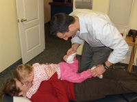
| Picture 1: A heel to buttocks test is displayed on a child. If the right leg produces a greater resistance compared to the left leg, during flexion towards the buttocks, then this would indicate a right posterior sacral base. |
||
 |
||
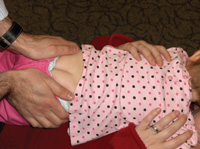
| Picture 2: A sacral squeeze test is performed on a child. If the crease that forms deviates to one side, this would indicate that the sacral base has subluxated anterior on the ipsilateral side of the deviation. The test in this picture displays a normal finding. |
||
 |
||
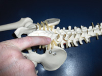
| Picture 3: Contact for a posterior base subluxation is displayed on a skeletal model. Note how a pinkie contact is utilized. |
||
 |
||
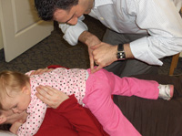
| Picture 4: Contact for a right posterior base subluxation is displayed on a child. Note how the pinkie contact is reinforced with the stabilization hand. |
||
 |
||
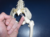
| Picture 5: Contact for an anterior inferior sacral base is displayed on a skeletal model. Note how the pinkie contact is on the caudal aspect of the sacro-tuberous ligament. |
||
 |
||
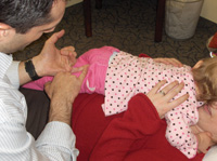
| Picture 6: Contact for an anterior inferior sacral base is displayed on a child. Note how the pinkie contact is stabilized, and that the sacro-tuberous ligament contact is on the ipsilateral side of the anteriority. Advertisement
|
CASE STUDY:
A mother and father bring their 11-month-old daughter to the clinic. The parents inform the doctor that the child is a happy and healthy child. However, they are concerned that since the child has been trying to stand and walk more and more, she has been having frequent falls onto her buttocks. The parents also inform the doctor that the child will utilize a piece of furniture or the parent’s leg to pull herself to a standing position, attempt one or two steps, and suddenly lose her balance and fall onto her buttocks. This process occurs several times per day, so due to this repetitive falling, the parents would like to have the child’s spine assessed. Overall assessment of the child’s spine reveals that the child is subluxation free, from the occiput to the lumbar spine. However, static and motion palpation of the pelvis reveals that the right sacro-iliac joint produces squirming in the child upon palpation, and that the sacral base lacks joint play when assessing posterior to anterior motion. All other analyses are unremarkable.
This case scenario is typical of a child presenting with a sacral subluxation. However, how does a chiropractor confirm if the sacrum has subluxated anterior or posterior, as palpation alone cannot fully determine the subluxation pattern? In this edition of Technique Toolbox, I will discuss the pediatric sacrum, how to confirm if the sacral base has subluxated posterior or anterior, and how to adjust these two separate pediatric subluxations.
THE PEDIATRIC SACRUM
The pediatric patient’s pelvis is a primary area of subluxation, just as in the adult. However, there is a major difference between subluxations that occur in the sacrum of a child and those that occur in an adult. Since the child’s sacrum is not completely fused, and its articulations with the ilium are not fully established, the pediatric patient’s sacral base can bilaterally and ipsilaterally subluxate in either the anterior or posterior direction. Therefore, in a child, the doctor must differentiate between an anterior or posterior subluxated sacral base, as palpation alone does not provide enough data for the chiropractor.
Step 1: Analysis for Posterior Sacral Base (See picture 1)
Doctor will perform a “Heel to Buttocks” test to determine if the sacral base has subluxated posteriorly.
- Patient: Prone (if the child is an infant, place the infant within a pediatric cushion, or have the mother lie supine and the infant can lie prone on mother’s chest and stomach).
- Doctor: Either side of patient.
- Flex one of the patient’s legs until the heel meets the buttocks. Repeat for the other leg.
- Normally, the patient’s heels will meet the buttocks pain free and with equal resistance on both sides. If this is the case, then the pediatric patient does not have a posterior sacral base subluxation.
- However, if one heel is not able to touch the buttock, produces an increased resistance in comparison to the opposite side, or produces pain during the test, this would indicate that the sacral base has subluxated posteriorly.
Step 2: Analysis for Anterior Sacral Base (See picture 2)
Doctor will perform a “Sacral Squeeze” test to determine if the sacral base has subluxated anteriorly.
- Patient: Prone (child placed within a pediatric cushion, or mother is supine and infant lies prone on mother’s chest and stomach).
- Doctor: Either side of patient.
- Squeeze the gluteal muscles together and observe the crease that forms midline, cephalad to the buttocks.
- If the sacrum is articulating normally, the crease formed should point straight cephalad.
- If the crease deviates to the left or right side, this will indicate that the sacrum has subluxated anterior and inferior, to the ipsilateral side of the deviation.
The analytical test that yields a positive result will determine which of the following adjustments will be performed on the child.
Step 3: Posterior Sacral Base Adjustment (See pictures 3 and 4)
- Patient: Prone.
- Doctor: Same side as lesion.
- Contact: Pinkie contact on lateral aspect of the sacral base on the affected side.
- Stabilize: Opposite pinkie finger stabilizing original contact.
- LOC: P-A. 2-6 ounces of pressure.
- Note: This adjustment can be performed using a drop piece, a toggle board or sustained contact pressure in the proper line of correction.
Step 4: Anterior Inferior Sacrum Correction (See pictures 5 and 6)
- Patient: Prone.
- Doctor: Same side as lesion.
- Contact: Pinkie finger under ipsilateral sacro-tuberous ligament.
- Stabilize: Opposite pinkie finger stabilizing original contact.
- LOC: L-M, I-S sustained contact. 2-6 ounces of pressure. Hold contact until tension subsides.
In this correction, the doctor is using the sacro-tuberous ligament as an anchor to set the sacrum back to its neutral position. This is the preferred adjustment for infants, because it insures patient comfort.
Assessing and correcting sacrum problems in our pediatric patients is essential. As a child learns to crawl and walk, the probability of subluxations in the pelvic area is increased. Therefore, it is critical that each child be assessed for subluxations and adjusted as indicated, to help prevent future spinal problems. As usual, I have only scratched the surface with pediatric adjusting. If you would like to learn more about assessing and adjusting children, please visit www.icpa4kids.com. If you would like to see a specific technique in a future edition of Technique Toolbox, please contact me at johnminardi@hotmail.com.
Until next time . . . adjust with confidence! •
Print this page