
Technique Toolbox: Palmer Upper Cervical Specific
By John Minardi BHK DC
Features Clinical TechniquesSAMPLE CASE: A 40-year-old office worker presents to the clinic with headaches, back
pain, and the occasional shooting pain into the right buttock.
SAMPLE CASE
A 40-year-old office worker presents to the clinic with headaches, back pain, and the occasional shooting pain into the right buttock. The patient informs the doctor that he has recently been transferred to this neighbourhood, and has been looking for a local chiropractor to assist with his problem, which he has been dealing with, on an intermittent basis, for the past 10 years. He has always found visits to a chiropractor extremely helpful. The patient relays that he has been subjected to long working hours, of late, and is spending much of his time focused on his computer screen. He attributes his issues to this. Physical examination reveals a palpatory left lateral atlas subluxation, a compensatory right posterior ilium, and hypertonic piriformis on the right. X-ray analysis of the cervical spine reinforces the palpatory atlas finding, and S-EMG analysis also reveals a thermal deviation at C1 on the left. All other radiological and neurological examinations are unremarkable.
During the report of findings, the doctor explains the subluxation findings to the patient before proceeding with a plan of management for the case. At the end of the report, the patient asks the doctor if he is proficient in upper cervical adjusting, as this method was used on him in the past and is what he prefers to be adjusted with. This particular doctor is trained in Palmer Upper Cervical Specific Technique, also known as “Hole in One” (HIO), and informs the patient that he can certainly accommodate his request.
What is the Hole in One technique? Who originally developed this technique and how is it utilized? In this edition of Technique Toolbox, I will answer these questions and more.
HISTORY
HIO technique was developed by B.J. Palmer following his extensive clinical research conducted at the Palmer Research Facility. Prior to 1931, B.J. Palmer felt that a subluxation could be located at any vertebra in the spine, including the sacrum and coccyx. However, from the very beginning, he felt that most subluxations occurred in the upper cervical spine, specifically at the atlas and axis vertebrae. At that time, Palmer defined vertebral subluxations to include the following criteria:1
- misalignments of the vertebra in relation to adjacent segments;
- occlusion of a foramen or spinal canal;
- pressure or tension on the spinal nerves or spinal cord;
- interference of transmission of mental impulses.
Palmer also made the clear distinction that chiropractors do not treat the diseases of patients, but rather, chiropractic care was focused on adjusting vertebral subluxations.
In 1931, the HIO technique was revealed to the chiropractic profession by B.J. Palmer. Palmer described that an adjustment, “with that extra something,” releases interference in one area, without creating more at other areas, therefore, creating a “Hole in One.” Palmer’s intent was to create an adjustment that had maximum effect on mental impulses and was sustained for longer periods of time.
However, his presentation was greeted with tremendous controversy as well as acclaim. The controversy surrounding HIO appeared when Palmer declared that the only region of the spine in which a subluxation could occur was in the upper cervical spine. This revolutionary technique focused solely on an upper cervical subluxation and utilized a ”toggle-recoil” thrust for correction.
This Palmer Upper Cervical Specific Technique, and the concurrent philosophy that only the upper cervical spine could subluxate, remained at Palmer School of Chiropractic until the 1950s. After this time, segmental adjusting of the entire spine would be re-introduced.1
So, how does a chiropractor, today, utilize this classic technique?
 |
|
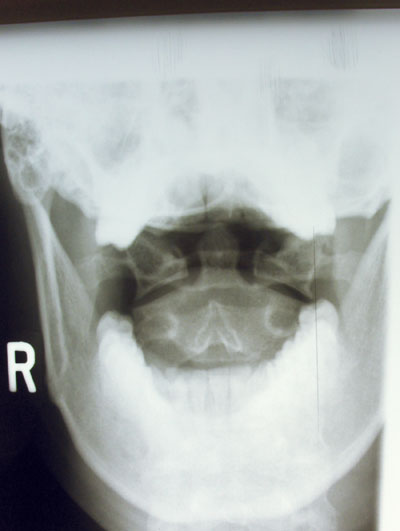
| Photo 1: An A-P open mouth cervical view is displayed. The markings note atlas subluxating to the patient’s left side. | |
 |
|
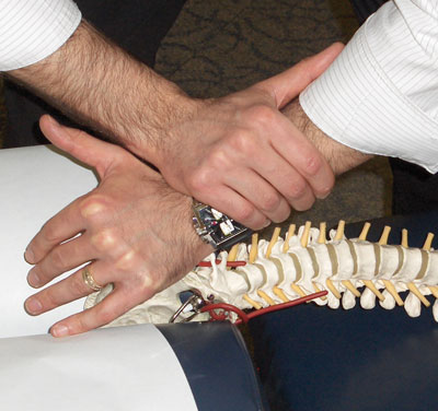
| Photo 2: HIO Toggle setup is displayed on the skeletal model. Note how a pisiform contact is on the lateral tip of the TVP of atlas, and the stabilization hand is in the anatomical snuff box of the contact hand. |
|
 |
|
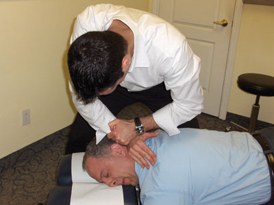
| Photo 3: HIO Toggle setup is displayed while the patient is in a prone position. Note how the patient’s head is turned so that the atlas laterality side is up. This setup can be used on a flat bench or knee-chest table. |
|
 |
|
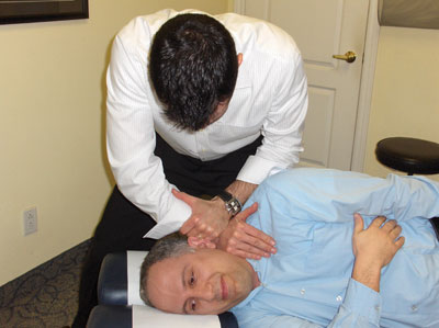
| Photo 4: HIO Toggle setup is displayed while the patient is in a side posture postion. Note that the cervical drop-piece may or may not be utilized in this position. | |
 |
|
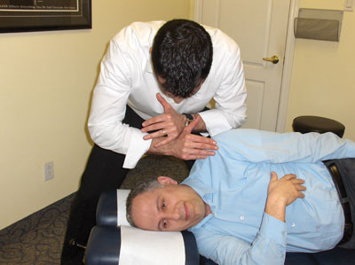
| Photo 5: Toggle recoil finish position is displayed. Note that the HIO toggle recoil adjustment begins with a thrust into the subluxated atlas, followed immediately by a recoil of the doctor’s contacts, as displayed in this photo. |
STEP 1: ANALYSIS
How classic HIO practitioners found the problem
First, the neurocalometer, a thermal device used to detect a change in heat, was used to locate the atlas subluxation. When the neurocalometer detected a thermal change in the skin – typically an increase in temperature – the device’s indicator would deviate to the left or right. This deviation represented a disruption of neural flow, indicating that a subluxation was present in that area. If the indicator deviated left, the subluxation was causing greater heat emission on the left side. (Current practitioners may utilize the S-EMG for a similar purpose.) However, this did not accurately describe the exact orientation of the subluxated atlas. Therefore, following the neurocalometer finding, X-ray analysis was utilized to confirm the precise direction of the subluxation.
Second, as I mentioned, specific X-ray analysis was utilized (see photo 1):
- A-P open mouth cervical view: Determines the side of laterality of the atlas.
- Draw vertical lines from the superior tip of the lateral mass bilaterally.
- Draw vertical lines from the inferior tips of the mastoid processes bilaterally.
- The side in which the lines converge indicates the side of laterality.
- Atlas rotation was typically measured on the base posterior view.
It should be noted that HIO X-ray analysis has certainly evolved over time, and may involve other aspects, which are out of the scope of this article.
STEP 2: CORRECTION USING AN HIO TOGGLE RECOIL ADJUSTMENT (See photos 2-5)
Patient:
- On knee-chest table: Kneeling. Chest prone on table. Head rotated with atlas laterality side up.
- On flat-bench: Prone, with head rotated with atlas laterality side up.
- Or, side posture, with atlas laterality side up. Cervical piece remains neutral, and is raised to accommodate the patient’s shoulder width.
Doctor: Side of the table
- Contact: Pisiform contact on the lateral tip of the C1 TVP.
- Stabilization hand: Anatomical snuff box of contact hand.
- LOC: L-M thrust with an added recoil following the thrust. Can be used with or without a drop-piece.
To say that B.J. Palmer was a leader and visionary for chiropractic would be an enormous understatement. With his enthusiasm, dedication and unwavering principles, undoubtedly he developed and propelled the growth of our profession further than anyone else in the history of chiropractic. An individual chiropractor may not agree with everything that B.J. Palmer did in his lifetime of chiropractic service, but one must never forget that without B.J. Palmer, they would not have the opportunity to be a chiropractor today. Let us always be mindful and respectful of the original visionary of chiropractic, and the technique that he developed. As usual, I have only scratched the surface of HIO.
If you would like to learn more, please go to www.upcspine.com. If you have any questions or comments, you can reach me at johnminardi@hotmail.com.
Until next time . . . Adjust with Confidence!
Dr. John Minardi is a 2001 graduate of Canadian Memorial Chiropractic
College. A Thompson-certified practitioner and instructor, he is the
creator of the Thompson Technique Seminar Series and author of The
Complete Thompson Textbook – Minardi Integrated Systems. In addition to
his busy lecture schedule, Dr. Minardi operates a successful private
practice in Oakville, Ontario. E-mail: johnminardi@hotmail.com, or visit
www.ThompsonChiropracticTechnique.com.
Print this page