Now, on to today’s case. Many thanks to Dr. Paul Kennedy of Toronto for sharing this dramatic case.
 |
|
| Figure 1
|
May you, my friends and colleagues, and your families, have a safe and most enjoyable holiday season. I would like to take this opportunity to thank Dr. Bill Mulhall for organizing my Radiology Rounds Seminar for the Manitoba Chiropractors’ Association in their beautiful new facility, and Dr. Salima Ismail, president of the Eastern Ontario Chiropractic Society for her hard work in putting together my Radiology Rounds Seminar in Ottawa last month. It was very enjoyable to meet and interact with all of the doctors in Winnipeg and Ottawa.
Now, on to today’s case. Many thanks to Dr. Paul Kennedy of Toronto for sharing this dramatic case.
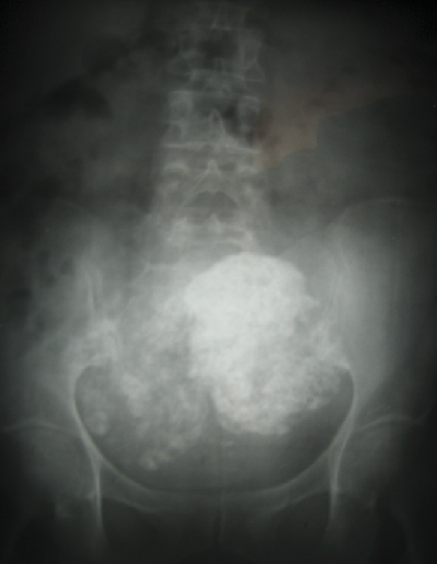
This 69-year-old woman presented with chronic, dull low back pain and abdominal fullness. Radiographic examination (Figure 1) revealed two large spheres of mottled or flocculent calcification in the pelvic inlet; the first measuring approximately 95mm x 110mm and the second measuring 85mm x 100mm.
Discussion
Uterine fibroids (fibroma, leiomyoma) are benign tumors which grow from the muscle layers of the uterus. They are the most common benign neoplasm in females, and may affect about 25 per cent of white and 50 per cent of black women during the reproductive years. Uterine fibroids often do not require treatment, but when they are problematic, they may be treated surgically or with medication — possible interventions include a hysterectomy or hormonal therapy.
As women age, they are more likely to have uterine fibroids, especially from their thirties and forties until menopause. About 80 per cent of women have uterine fibroids by the time they reach age 50. Most have mild or no symptoms. But fibroids can cause serious problems that need treatment.
The cause of uterine fibroids is not known. But after fibroids develop, the hormones estrogen and progesterone appear to influence their growth. After menopause, when hormone levels decline, fibroids often shrink or disappear. In very rare cases, malignant growths on the smooth muscles inside the womb can develop, called leiomyosarcoma of the womb.
Pathology and histology
Leiomyomas grossly appear as round, well circumscribed (but not encapsulated), solid nodules that are white or tan, and whorled. The size varies, from microscopic to lesions of considerable size. Typically, lesions the size of a grapefruit or bigger are felt by the patient herself through the abdominal wall. Leiomyomas are estrogen sensitive and have estrogen receptors. They may enlarge rapidly during pregnancy due to increased estrogen levels. Fibroids tend to regress following menopause because of lowered levels of estrogen. Hormonal therapy is based on this concept.
Symptoms
The symptoms depend on the size, location, number, and the pathological findings. Fibroids, particularly when small, may be entirely asymptomatic and found incidentally on plain film. Important symptoms include abnormal gynecologic hemorrhage, heavy or painful periods, abdominal discomfort or bloating, back ache, urinary frequency or retention, and in some cases, infertility. During pregnancy, they may be the cause of miscarriage, bleeding, premature labour, or interference with the position of the fetus.
Diagnosis
Often found incidentally on plain film when calcified, diagnosis is usually accomplished by bimanual examination, better yet by ultrasound. Sonography will depict the fibroids as focal masses with a heterogeneous texture, which usually cause shadowing of the ultrasound beam. Magnetic resonance imaging (MRI) can be used to define the depiction of the size and location of the fibroids within the uterus. No imaging modality can clearly distinguish between the benign uterine leiomyoma and the malignant uterine leiomyosarcoma; however, the latter is very rare, and there is a tremendous prevalence of the former. For this reason, biopsy is rarely performed and if performed, is rarely diagnostic. Should there be an uncertain diagnosis after ultrasounds and MRI imaging, or should there be questions regarding whether the fibroid is interfering with fertility, a laparoscopy is one option for further information to be gathered regarding the exact size and location of the fibroid.
Treatment
The presence of fibroids does not mean that they need to be treated; this is dependant on the symptomatology and presence of related conditions. The presence of uterine fibroids can cause problems which can be corrected with surgery – hysterectomy (uterine removal) or myomectomy (fibroid removal). Although a myomectomy cannot prevent the recurrence of fibroids at a later date, such surgery is increasingly recommended, especially for women who have not completed bearing children, or who express an explicit desire to retain the uterus.
Malignancy
Very few lesions are, or become, malignant. Signs that a fibroid may be malignant are rapid growth or growth after menopause. Such lesions are typically a leiomyosarcoma on histology. There is no consensus, among pathologists, regarding the transformation of leiomyoma into a sarcoma. Most pathologists believe that a leiomyosarcoma is a de novo disease. •
Print this page