This 13-year-old hockey player presented with a painful left knee that
clicked a lot on flexion and extension. His mother had reported that a
“benign bone growth” had been surgically removed a few years earlier.
 |
||
| Figure A
|
||
 |
||
| Figure B Advertisement
|
Thank you to my good friend, Dr. Peter Amlinger, of Mississauga, Ontario, for this case.
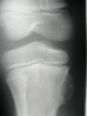
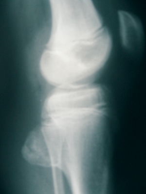
This 13-year-old hockey player presented with a painful left knee that clicked a lot on flexion and extension. His mother had reported that a “benign bone growth” had been surgically removed a few years earlier. Examination revealed subluxations at atlas and at the sacroiliac joints. The left hamstring was extremely tight and tender to palpation along the medial aspect (semimembranosis). There was a noticeable click from this area as the patient extended his knee. Palpation revealed a large hard bulbous mass projecting from the posteromedial border of his left proximal tibia. Figures A and B demonstrate the well-defined exostosis at the posteromedial aspect of the proximal tibial metaphysis.
Upper cervical specific adjustments were administered as needed, beginning with a frequency of three times a week. After 12 atlas adjustments, the pain and click were gone and the hamstring length had normalized. The patient was referred for an orthopedic consultation with respect to the recurrence of the osteochondroma.
OSTEOCHONDROMA Discussion:
- osseous projection, often with cartilaginous cap arising from host bone cortex
- develops slowly during childhood and adolescence
- cortex continuous with cortex of host bone, and medullary portion continuous with central spongiosa
- cessation of tumour development at skeletal maturity
- most frequent complaint is a painless mass
- may be asymptomatic, or symptoms may be produced by the mechanical pressure of the tumour on contiguous vascular or neurological tissue
- symptoms may follow mild trauma or fracture of the lesion
- most common location is tubular bone near metaphysis; usually knee
- can be pedunculated (with stalk) or sessile (with wide base)
- can be solitary (1 per cent malignant potential) or multiple (hereditary multiple exostosis; 20 per cent malignant potential)
Print this page