This case is courtesy of the radiology department at Southern
California University of Health Sciences. This 22-year-old woman
presented to the outpatient clinic complaining of diffuse mid-back
pain, right shoulder pain, and occasional bouts of non-exertional
shortness of breath.
This case is courtesy of the radiology department at Southern California University of Health Sciences. This 22-year-old woman presented to the outpatient clinic complaining of diffuse mid-back pain, right shoulder pain, and occasional bouts of non-exertional shortness of breath.
She had received relief with chiropractic care, when the pain first began a year previously, but, in the past week, she had suffered a recurrence of the symptoms.
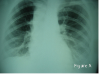
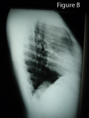
Physical examination revealed a soft tissue mass just above the right clavicle. Significant findings on X-ray examination included widening of the upper mediastinum with an extra-pleural sign (Figure A) and opacification of the anterior clear space (Figure B).
 |

|
|
| Figure A | Figure B |
Diagnosis:
Hodgkin’s Lymphoma
Discussion:
The Extra-pleural Sign and Opacification of the Anterior Clear Space
Lesions arising in structures which border the extra-pleural space may project into it. Examples include rib lesions (e.g., metastasis), soft tissue neoplasia, infection – especially tuberculosis – and chest wall hematoma. These lesions in the extra-pleural space often produce a characteristic X-ray appearance. Because of the intact layers of parietal and visceral pleura overlying it, an extra-pleural lesion often presents a very sharp, curvilinear contour facing the lung. Additionally, the superior and inferior edges of an extra-pleural lesion are usually tapered due to the difficulty in the lesion attempting to strip the pleura away from the chest wall. This sharp, tapered contour, as seen in Figure A, are hallmarks of the “extra-pleural sign.” The third classic indicator of an extra-pleural lesion is that the lesion’s margin is convex toward the lung. This is also seen in Figure A. In this case, the lesion is mediastinal, and, additionally, has opacified the usually relatively radiolucent space normally seen above the cardiac silhouette on the lateral projection. Extra-pleural lesions usually do not break into the pleural space – therefore, pleural effusion and thickening are generally not present.
When I first studied chest radiology at CMCC, my professor at the time, Dr. Lindsay Rowe, taught me that the superior clear space is opacified most commonly by one of four conditions in the mnemonic “3 T’s and an H”, namely, thyroid (goiter), teratoma (thymoma), tumours of the sternum (mostly metastasis), and Hodgkin’s lymphoma.
Hodgkin’s disease is what was proven, on biopsy, in this particular lady.
Reference:
Felson B, et al. Principles of Chest Roentgenology: a Programmed Text. 1965, WB Saunders, Philadelphia, PA.
A 1983 CMCC graduate, Marshall Deltoff Marshall Deltoff, DC, DACBR, FCCR(C), completed his radiology residency at Los Angeles College of Chiropractic. He is a past radiology department chairman and residency coordinator at CMCC, and he initiated the radiology curriculum at UQTR. Dr. Deltoff has lectured throughout North America, and is co-author, along with Dr. Peter Kogon, DACBR, of the radiology text “The Portable Skeletal X-ray Library” published by Mosby-Yearbook of St. Louis. Dr. Deltoff can be reached at:
Images Radiology Consultants,
16 York Mills Road,
Toronto, Ont. M2P 2E5
Tel: (416) 512-2225
Fax: (416) 512-2226
e-mail: marshdel@rogers.com
Print this page